Physics of the Tympanogram

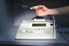
The tympanometer measures the “admittance” or “compliance” of the tympanic membrane while different pressures are being applied to the external ear canal. The compliance of the TM is measured in cubic centimeters, and the pressure in the ear canal is measured in decapascals (daPa). The probe has different size “plugs” that provide a seal at the entrance to the external ear canal. The tip of the probe has a pressure transducer that changes the pressure in the external ear canal from negative, through atmospheric pressure, to positive pressure. While the pressure is changing, a sound transmitter sends a sound wave to the tympanic membrane. The wave that is reflected from the TM is then picked up by a microphone in the probe. The tympanometer measures the energy of the reflected sound.
If the middle ear space is filled with fluid, most of the sound is reflected back to the probe from the stiff tympanic membrane. If the middle ear space is filled with air, and the ossicles are intact, energy is absorbed by the tympanic membrane, ossicles, and inner ear structures. The tracing will read “normal”. If there is disruption of the ossicles, or if a portion of the TM is flaccid, a large amount of energy will be absorbed into the TM and the tracing will display an abnormally peaked compliance.
Children under six months of age may demonstrate falsely increased compliance, due to the fact that the ear canal skin and cartilage is quite lax. In this situation, the child may have a middle ear filled with fluid, but still register a compliance within the normal range.
The tympanometer automatically calculates the canal volume. If there is cerumen or other material occluding the ear canal, the volume will measure abnormally low. An accurate tracing cannot be obtained until this material is removed.
Similarly, if there is a perforation of the tympanic membrane the tympanometer will measure an unusually large canal volume, because the space of the middle ear and mastoid air cells will be included in the volume calculation.
Normal Tympanogram
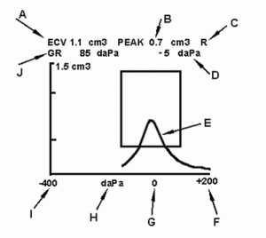
View a normal tympanogram and it’s elements below:

A – ECV = External Canal Volume. This varies from 0.5 to 1.2 cc. A smaller value would indicate that a foreign body, cerumen, or other material is occupying some space in the external canal.
Alternatively, a small volume might simply represent poor positioning of the probe. A large canal volume indicates that there may be a perforation of pressure equalizing tube in the tympanic membrane.
B – Peak = This is the peak compliance of the tympanic membrane measured in cc.
The y axis marks the level of compliance.
Note that this tracing will measure compliance accurately up to 1.5 cc.
C – R = right. This is a tracing of the right ear.
D – daPa = decapascals. This is the pressure at which peak is located.
These pressures read on the x axis.
E – Pressure tracing = This tracing records the compliance of the tympanic membrane at a continuous sweep from negative to atmospheric to positive pressure applied to the ear canal.
F – +200 = indicates 200 decapascals of positive pressure in the ear canal.
G – 0 = indicates zero (atmospheric) pressure in the ear canal.
H – daPa = decapascals is the unit of measure of pressure in the external ear canal.
I – -400 = negative air pressure in the external ear canal.
J – GR (Gradient) = A measure of the shape (width) of the tracing.
A gradient greater than 150 daPa is often associated with a collection of fluid in the middle ear space.
The combination of a large gradient (>150 daPa) and low compliance (<0.2 cc) is associated with a 95% or greater likelihood of a middle ear effusion.
Classifying Tympanograms
Tympanograms are classified, for ease of discussion, by the following rules:
| Type | Description |
|---|---|
| A | Peak compliance > 0.2 ml near 0 daPa middle ear pressure (MEP) |
| A’ | Peak > 0.2 ml MEP -10 daPa to -99 |
| A+ | Peak > 0.2 ml MEP > +50 daPa |
| As | Peak < 0.2 ml MEP -99 daPa to +49 daPa |
| C1 | Regardless of Peak MEP -100 daPa to -150 daPa |
| C2 | Regardless of Peak MEP -151 daPa to -200 daPa |
| C3 | Regardless of Peak MEP < -201 daPa |
| B | No Peak anywhere on the tracing |
Abnormal Tympanograms
Read the cases below and think about how the tympanogram would be affected. When you are done, display the tympanogram for each case to see how close you came.
A two-year-old boy who comes to the office for a well-child visit. Mother thinks the child is not hearing as well as he should.
Examination of the tympanic membranes reveals normal color and mobility on pneumatic otoscopy. You check a tympanogram to assure yourself that no fluid is present in the middle ear.
A one-year-old girl with bilateral pressure equalizing tubes. She has had recurrent infection in the left ear, and you suspect that the pressure equalizing tube may be obstructed.
You check a tympanogram, paying particular attention to the shape of the curve and the external canal volume.
An eight-month-old boy with acute onset of pain in the right ear.
The ear appears red and you have some difficulty defining the structures.
A three-year old girl who comes back for a recheck on her right ear, six weeks following an acute ear infection.
The tympanic membrane is dull and non-erythematous. The landmarks are easily visualized.
