Secondary lesions may evolve from primary lesions, or may be caused by external forces such as scratching, trauma, infection, or the healing process. The distinction between a primary and secondary lesion is not always clear.
Click any of the images below to view them in a higher resolution.
This is an accordion element with a series of buttons that open and close related content panels.
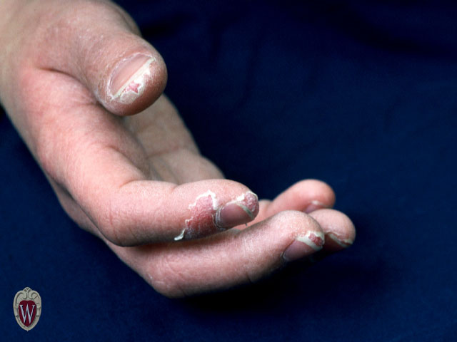
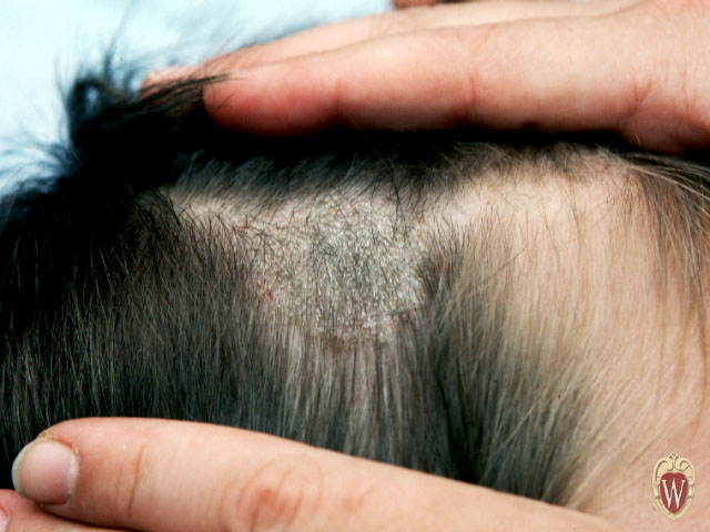
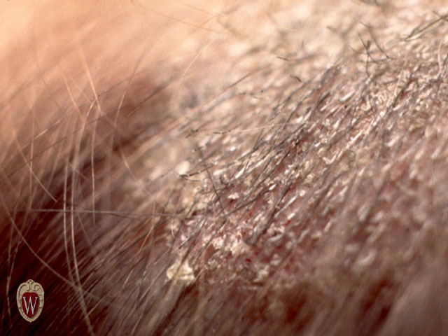
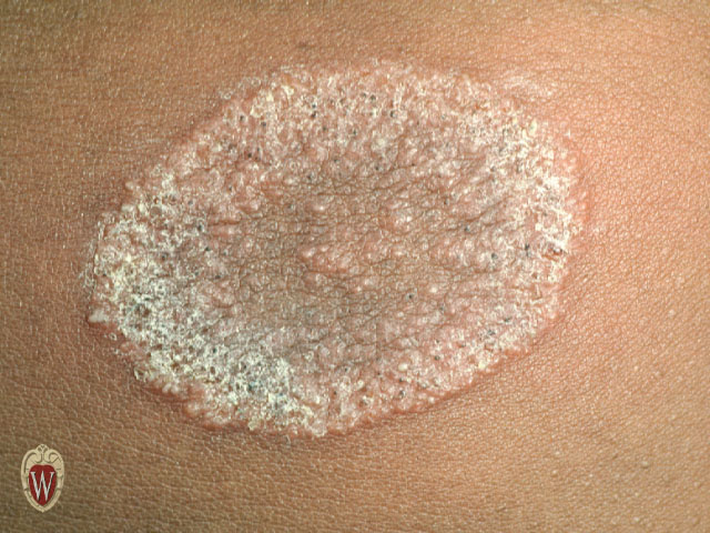
Scale
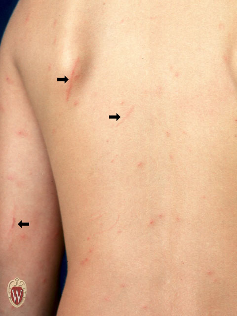
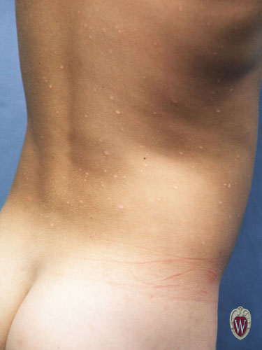
A macule is a change in the color of the skin. It is flat, if you were to close your eyes and run your fingers over the surface of a purely macular lesion, you could not detect it. A macule greater than 1 cm. may be referred to as a patch.
|
|
|
|
|
|
|
|
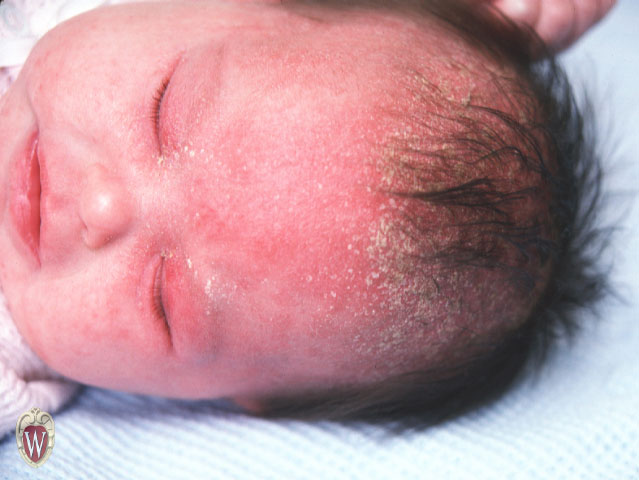
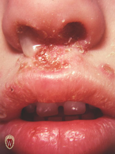
Crust
Crusting is the result of the drying of plasma or exudate on the skin. Please remember that crusting is different from scaling. The two terms refer to different phenomena and are not interchangeable. One can usually be distinguished from the other by appearance alone.
|
|
Atrophy
Atrophy is thinning or absence of the epidermis or subcutaneous fat.
|
|
|
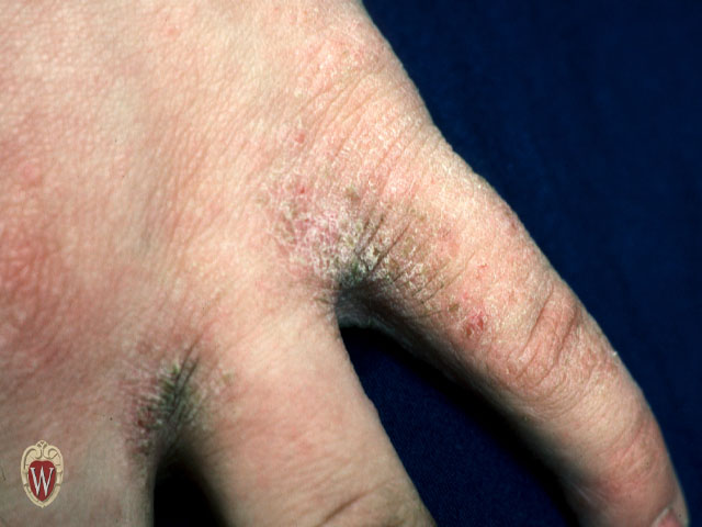
Lichenification
“Lichenification” refers to a thickening of the epidermis seen with exaggeration of normal skin lines. It is usually due to chronic rubbing or scratching of an area.
|
|
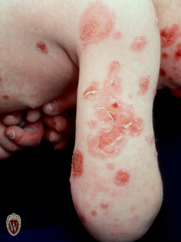
Erosions
Erosions are slightly depressed areas of skin in which part or all of the epidermis has been lost.
|
|
|
Excoriation
Excoriations are traumatized or abraded skin caused by scratching or rubbing.
|
|
Fissure
A fissure is linear cleavage of skin which extends into the dermis.
Erosions are slightly depressed areas of skin in which part or all of the epidermis has been lost.
|
|
|

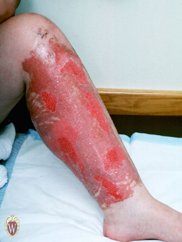
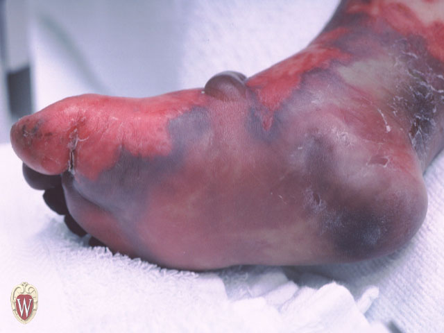
Ulceration
Ulcerations occur when there is necrosis of the epidermis and dermis and sometimes of the underlying subcutaneous tissue.
|
|
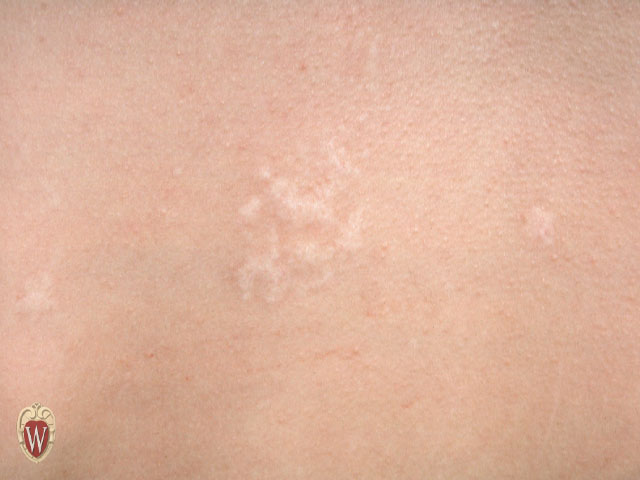
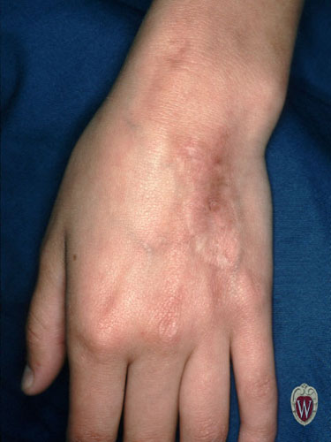
Scar
Scars are the permanent fibrotic changes that occur on the skin following damage to the dermis. Scars may have secondary pigment characteristics.
|
|
|
|
|
|
|
|
Eschar
An eschar is a hard plaque covering an ulcer implying extensive tissue necrosis, infarcts, deep burns, or gangrene.
|
|
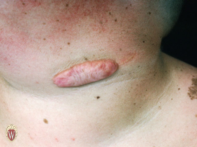
Keloids
Keloids are an exaggerated connective tissue response of injured skin that extend beyond the edges of the original wound.
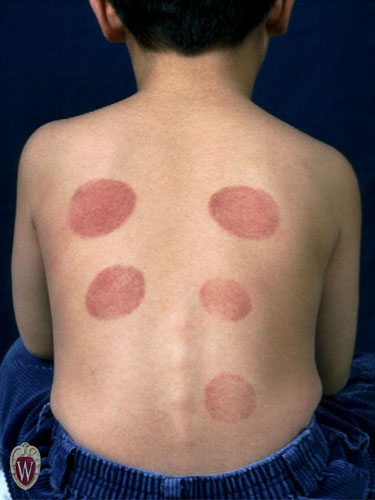
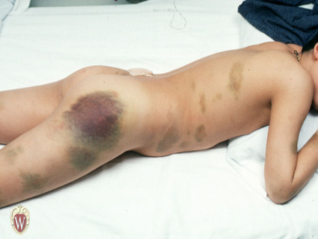
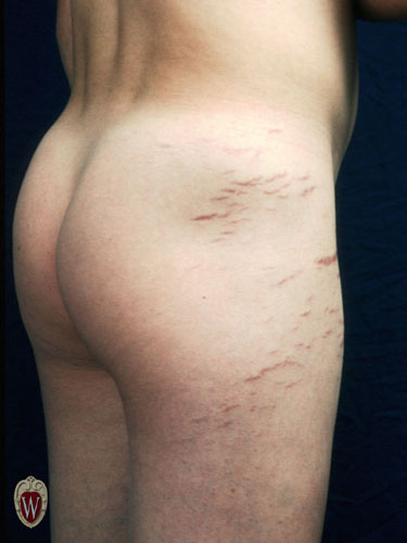
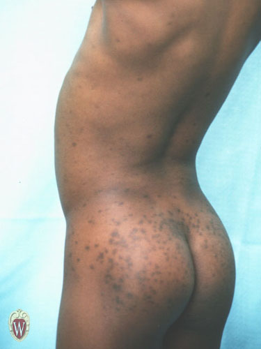
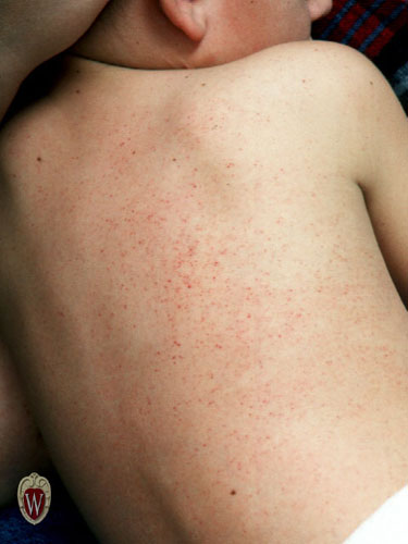
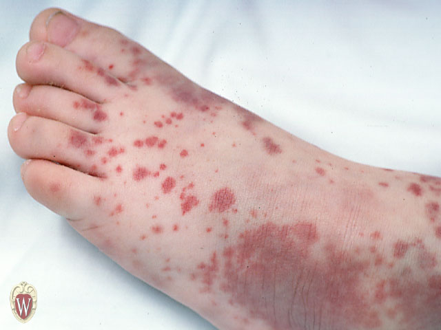
Petechiae, Purpura, and Ecchymoses
Three terms that refer to bleeding that occurs in the skin are petechiae, purpura, and ecchymoses. Generally, the term “petechiae” refers to smaller lesions. “Purpura” and “ecchymoses” are terms that refer to larger lesions. In certain situations purpura may be palpable. In all situations, petechiae, ecchymoses, and purpura do not blanch when pressed. If there is any question, press on the lesions carefully with a glass slide. Don’t break the slide or cut the patient.
|
|
|
|
|
|